Hip pain can stem from many causes including problems with a patient’s bony structures as well as in their soft tissues. A correct diagnosis is critical to ensuring that the best possible treatment plan is chosen. In fact, a delay in making the right diagnosis can have adverse consequences like arthritis or even the need for surgery that may have been avoided otherwise.
MRI (magnetic resonance imaging) is the most sensitive test for evaluating the bones and surrounding soft tissues.
The first step in obtaining an accurate diagnosis is always a complete medical history and physical exam by a physician who has experience in this area. This information helps guide what type of imaging study could be helpful. Usually, traditional x-rays are a good place to start because these can show fractures or arthritic changes. If your x-rays are normal and you have persistent hip symptoms, your doctor may choose to proceed with an MRI.
MRI (magnetic resonance imaging) is the most sensitive test for evaluating the bones and surrounding soft tissues. A MRI can pick up stress fractures or even bone bruises that a plain x-ray will usually miss. It can also detect the early findings of arthritis, even when the x-rays are normal, because it can show changes in your cartilage and the underlying bone.
An MRI is a good tool for evaluating the many causes of pain that may surround the hip joint itself. There are several tendons that insert around the hip and that can become inflamed or degenerated. Bursitis, usually located at the outside (lateral) part of the hip, can be painful. In addition, if you have had a recent injury or engaged in excessive athletic activity, your muscles can become injured (known as a “muscle strain”) and this can be detected by MRI.
An MRI will often show unexpected causes of hip pain that may be originating from other nearby structures like the sacroiliac joints, pubic bones, or even the lower lumbar spine. More worrisome sources of pain that could be coming from tumors, an infection or necrosis of the bone (also known as AVN) can also be eliminated from the list of potential causes by doing an MRI. Some of these causes are evaluated by MRI with intravenous contrast.
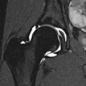
A common cause of hip pain, especially in younger patients, is a tear of the acetabular labrum, a soft tissue structure that lines the socket part of the hip joint. This can cause pain or a sensation of catching or popping in the joint. This type of injury is often best evaluated with a special type of MRI called an MRI Arthrogram. During this procedure, a radiologist injects fluid into the joint to get a better picture of the labrum.
Equal to getting a good quality MRI, it is important that an experienced radiologist – who is trained and specialized in orthopedic and sports imaging – reads your study. These doctors, known as musculoskeletal radiologists (MSK), are skilled at identifying subtleties that can be missed if an MRI is read by a non-specialist or a less experienced radiologist.
Wake Radiology is proud to have a team of seven subspecialty trained MSK radiologists who are dedicated to orthopedic and sports imaging and we invite you to learn more about hip-related imaging procedures. And, if you and your provider determine you need a hip MRI, consider our West Raleigh office where we have dedicated orthopedic and sport imaging radiologists on staff every day.